Skip to content

DWRX2008 · SGLT2 inhibitor eyedrop for DR & DME

A drop in front. Drug at the back. No needle in the eye.

FIRST-IN-CLASSThe first SGLT2 inhibitor eyedrop

DWRX2008 is a first-in-class SGLT2 inhibitor eyedrop: a 2% nano-micelle solution of enavogliflozin, a sub-nanomolar SGLT2 inhibitor. It skips the cornea and travels around the eye — through the conjunctiva and sclera — to reach the choroid and retina, where diabetic retinopathy and macular edema start.

Co-developed byDaewoong TherapeuticsDaewoong PharmaceuticalHanAll Biopharma

Class
1stSGLT2 inhibitor developed as an eyedrop
SGLT2 potency
0.43 nMSGLT2 IC50 of enavogliflozin
Efficacy
≈ IVTLeakage cut on par with Lucentis® and Eylea®
How a DWRX2008 eyedrop reaches the retina An eye in cross-section. A drop lands on the front of the eye; drug-loaded nano-micelles travel along the conjunctiva and sclera around the globe and pass through the choroid into the retina at the back of the eye, around the macula. Only a trace goes through the cornea, and none is found in the lens or vitreous. TRANS-CONJUNCTIVAL ROUTE 2% CONJUNCTIVA SCLERA CHOROID RETINA MACULA OPTIC NERVE VITREOUS CORNEA <0.001% TO RETINA LENS ENAVOGLIFLOZIN NANO-MICELLE LENS & VITREOUS: NOT DETECTED (QWBA)
  1. 01Drop on the eye
  2. 02Conjunctiva
  3. 03Sclera
  4. 04Choroid
  5. 05Retina

01The unmet need

Diabetic eye disease is treated with a needle in the eye.

Anti-VEGF injections into the vitreous work — but they are costly, invasive and repeated every one to four months, so many patients fall behind on treatment.

  1. 01

    Restricted to intravitreal injections

    Standard care for DR and DME is an anti-VEGF antibody injected into the eye, given in a clinic by a retina specialist.

  2. 02

    Cost and invasiveness hurt adherence

    Monthly to bi-monthly injections, each carrying risks of infection, raised IOP and retinal detachment, make patients drop out of care.

  3. 03

    Pills and drops don’t reach the retina

    Oral drugs rarely cross the blood–retinal barrier at safe doses; conventional eyedrops lose almost everything before the back of the eye.

Global prevalence · WHO

One in three people with diabetes develops retinopathy

Adults with diabetes

415 M→ 642 M

Diabetic retinopathy (35%)

145 M→ 224 M

Vision loss (11%)

45 M→ 70 M

TodayProjected

DR/DME is the leading cause of blindness in the U.S. adult workforce (≈4.1 M cases, ≈900 k vision-threatening). Diabetes is expected to reach ≈780 M people within 20 years.

Retinal disorder sales by indication · 2022

  • Wet AMD$4.3 B
  • Diabetic retinopathy$3.4 B
  • Macular edema after RVO$1.4 B

$9 B

Global VEGF-trap sales for angiogenic eye disease, 2022

95%

Share of retinal-disorder market value held by anti-VEGF injections

≈50%

Of that value taken by DR and DME

$14 B

DR market forecast by 2030 · CAGR 6.4%

Sources: WHO; IQVIA Global MIDAS 2023; Datamonitor 2018; Global Market Insights 2019; Research and Markets 2023.

02Why drops never got there

The retina sits behind every barrier the eye has.

A conventional drop aims straight through the cornea. Tears wash most of it away, and what crosses stalls in the front of the eye.

Conventional eyedrop

  1. Applied to the eye

    100%

  2. Lacrimal drainage & blinking

    most lost

  3. Across the cornea

    physical barriers

  4. Aqueous humor

    <3%

  5. Retina

    <0.001%

Oral tablet

  1. Swallowed

    systemic dose

  2. Whole-body circulation

    diluted

  3. Blood–retinal barrier

    tight junctions

  4. Retina (rabbit, 1–4 h)

    below LLOQ

  5. Higher dose?

    systemic limits

DWRX2008 drop

  1. Applied to the eye

    2% nano-micelle

  2. Conjunctiva

    highest level

  3. Sclera

    high level

  4. Choroid → retina

    reached

  5. Still at the retina

    detected at 48 h

Bars are illustrative; DWRX2008 levels are relative peaks from the rabbit QWBA study (log scale)

Conventional eyedrop figures: Worakul & Robinson (2000); Agarwal (2023); Wu (2023). Oral: comparative PK in NZW rabbits, below.

03How DWRX2008 gets there

A nano-micelle that takes the long way round.

The formulation largely bypasses trans-corneal absorption. Micelles cross the conjunctiva and sclera and diffuse inward through the choroid to the retina.

The carrier

HYDROPHILIC SHELLfaces tears & tissue HYDROPHOBIC COREholds the drug ENAVOGLIFLOZINpoorly soluble API
  • Amphiphilic

    Balanced with every ocular layer

    Better solubility and absorption; protects and stabilizes the encapsulated drug.

  • Particle size

    Deeper tissue penetration

    Longer retention in tissue for sustained release.

  • Flexible structure

    Slips through cell junctions

    Navigates narrow spaces with less risk of mechanical damage.

  • Surface charge

    Gentle on the eye

    Lower risk of irritation and inflammation; biocompatible with ocular tissue.

The route · peripheral ocular transport

Conjunctiva Sclera Suprachoroidal space Choroid Retina TRANS-CONJUNCTIVAL → TRANS-SCLERAL
  • Tailored formulation for far higher retinal delivery than conventional eyedrops.
  • A potent API is a must: enavogliflozin works in humans at under 1/30 the dose of leading SGLT2 inhibitors.

04Mechanism of action

Upstream of VEGF: treat the diabetic retina itself.

Anti-VEGF drugs block the last step. DWRX2008 acts where hyperglycemia first damages retinal vascular endothelial cells — at two points.

How hyperglycemia damages the retinal vasculature

  1. HG

    Hyperglycemia

    Chronically high glucose around retinal vascular endothelial cells and RPE.

  2. Glu

    SGLT2-mediated glucose uptake

    Glucose floods into cells through SGLT2, which human retinal cells express.

    ⊣ ① DWRX2008 blocks SGLT2
  3. ROS

    Mitochondrial dysfunction & excess ROS

    Overloaded mitochondria fragment and leak reactive oxygen species; AGE–RAGE signaling and inflammation reinforce the damage.

    ⊣ ② SIRT / AMPK / mTOR axis
  4. EC

    Endothelial dysfunction, poor barrier

    Capillaries leak and occlude in repeating cycles.

  5. VEGF

    VEGF ↑ · edema · neovascularization

    DME and proliferative DR — where today’s injections step in.

1

SGLT2-mediated: less glucose into the cell

Enavogliflozin blocks SGLT2 with an IC50 of 0.43 nM, cutting glucose uptake into retinal and vascular endothelial cells.

SGLT2 IC₅₀ 0.43 nM2-DG uptake −47%
2

Non-SGLT2: healthier mitochondria

Through the SIRT/AMPK/mTOR axis it lowers mitochondrial ROS, improves oxidative phosphorylation and fuel energetics, and supports mitochondrial biogenesis.

AMPK / SIRT inducerROS back to baseline

The result

Restored endothelial function, less VEGF

Barrier integrity recovers and high-glucose-driven VEGF secretion falls — a cause-level effect that can stand alone or sit beside anti-VEGF therapy.

Mechanism figure after Li (2022).

1stFIRST-IN-CLASS

The first SGLT2 inhibitor made to work as an eyedrop.

SGLT2 inhibitors are proven in diabetes, heart and kidney disease — but taken by mouth they do not reach the retina. DWRX2008 brings the class to the eye for the first time, with a mechanism no approved or late-stage retinal drug shares: it acts on the diabetic cause, not only on VEGF.

05Nonclinical evidence

From the cell to the fundus: five studies, one story.

Enavogliflozin protects human retinal cells; the drop puts it in the retina where the pill cannot; and in two disease models it cuts vascular leakage as much as an intravitreal antibody.

Study 1 · In vitro pharmacology

Less glucose in, less ROS, less VEGF

Human retinal pigment epithelial (RPE) cells express SGLT2. Under high glucose, enavogliflozin lowers glucose uptake, brings oxidative stress back to baseline and blunts the rise in VEGF secretion.

S1

Reduction of glucose uptake, ROS and VEGF in human retinal cells

Human RPE cell line · normal 5.5 mM vs high glucose 25 mM

Target is there

SGLT2 protein in human RPE cell lysates (western blot)

lysate 1lysate 2
SGLT2
β-actin
Glucose uptake

2-DG uptake, % of control, by enavogliflozin dose (nM)

−47%at 100 nM; dose-dependent from 0.16 nM
Oxidative stress

ROS level, % of control. High glucose raises ROS; enavogliflozin brings it down

157 → 106%ROS under high glucose, untreated vs 5 µM (p<0.001)
Neovascular signal

VEGF secretion, pg/mL. High-glucose-induced rise is suppressed

−18%VEGF vs high glucose alone at 1 µM, back to normal-glucose level

Limiting factors: DR/DME involves a complex interplay of ocular cells and pathogenic factors beyond high glucose. RPE is not the primary site of DR/DME pathology but was used for its availability. Significance as reported: * p<0.05, ** p<0.01, *** p<0.001 vs untreated control (glucose uptake) or vs high-glucose control (ROS, VEGF); ### p<0.001 vs normal glucose.
Study 2 · Comparative ocular PK

Same drug, same dose: only the drop reaches the retina

Rabbits received a single identical dose of enavogliflozin as DWRX2008 drops (both eyes) or as the oral tablet. Retinas were sampled at 1 and 4 hours. Every oral sample was below the quantitation limit.

S2

Oral enavogliflozin: insufficient retinal drug exposure

NZW rabbit · n = 6/group · n = 3/time point · single dose

G1 DWRX2008 ophthalmic solution · topical (R/L)G2 Enavogliflozin oral tabletDose identicalFrequency singleSampling retina, 1 h & 4 h
Was drug found in the retina?

One dot per animal and eye

Drug quantifiedBelow quantitation limit

Retinal exposure by route

Same drug, same dose, same time points

12 / 12retina samples with quantifiable drug after one DWRX2008 drop
0 / 12after the oral tablet — every sample below the quantitation limit

Oral SGLT2 inhibitors cannot cross the blood–retinal barrier at systemically safe doses; the eyedrop gets the drug there directly.

Study 3 · Ocular distribution (QWBA)

The drug goes around the eye — not through it

Quantitative whole-body autoradiography after a single [14C]-labelled drop. Levels are highest in the conjunctiva and sclera, the retina/choroid is exposed for 48 hours, and the lens and vitreous humor stay clear — the signature of a trans-conjunctival, trans-scleral route.

S3

DWRX2008: enough peripheral drug transport to the retina

NZW rabbit · n = 17 · single topical dose (R/L) · QWBA at 0.5, 1, 2, 4, 8, 24, 48 h · Charles River

[14C]enavogliflozin in ocular tissues

Relative level over 48 h, log scale. Tap a tissue to highlight it

Peak level by tissue

Relative peak level, outside → in

    48 hretina/choroid exposure still detected after a single drop
    • Undetectable drug in the lens and vitreous humor, yet clear exposure in the retina/choroid.
    • High concentrations in the conjunctiva and sclera point to a trans-conjunctival route of delivery.

    Curves redrawn from the study report figure as relative levels; absolute concentrations available under CDA.

    Study 4 · Efficacy in diabetic retinopathy

    A daily drop matches a Lucentis® injection

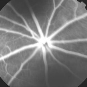
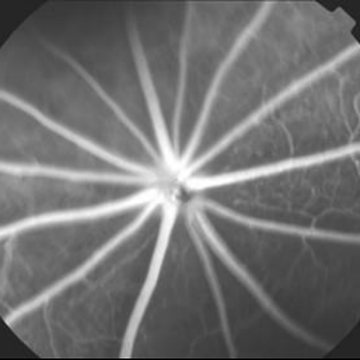
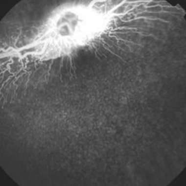
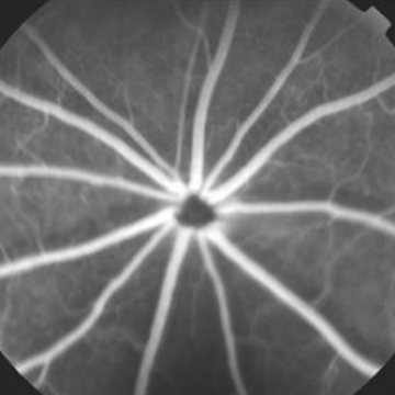
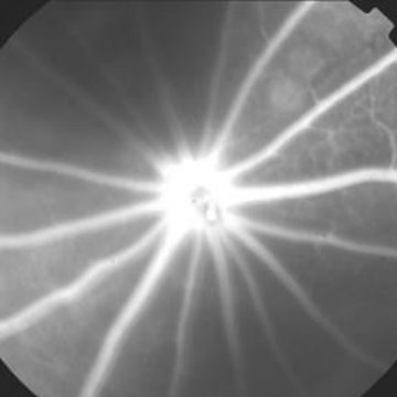
    In streptozotocin-induced diabetic Brown Norway rats, three weeks of once-daily DWRX2008 drops reduced retinal vascular leakage significantly versus vehicle — comparable to a single intravitreal injection of ranibizumab. Follow-up studies were run to find the minimum effective concentration.

    S4

    Suppression of vascular leakage in STZ-induced DR/DME rats

    Brown Norway rat · n = 10/group · STZ day 0 → final dose & FFA day 21

    GroupTreatmentDRDoseFrequencyRoute
    G1Normal controlN———
    G2VehicleY—QDTopical (R)
    G3Ranibizumab (Lucentis®)Y100 µg/eyeSingleIVT
    G4DWRX2008 ophthalmic solutionYUndisclosedQD × 21 dTopical (R)
    Fluorescein intensity, day 21

    % of normal; mean ± SD

    153 → 117%vehicle vs DWRX2008 (p<0.001); Lucentis® 120%
    Fundus fluorescein angiography, day 21

    Leakage shows as diffuse brightness around the vessels

    Fundus angiogram, normal rat
    G1Normal
    Fundus angiogram, diabetic rat with vehicle
    G2Vehicle
    Fundus angiogram, diabetic rat with Lucentis
    G3 · IVTLucentis®
    Fundus angiogram, diabetic rat with DWRX2008 drops
    G4 · DROPDWRX2008

    *** / * p<0.001 / p<0.05 vs G1 (normal). ### p<0.001 vs G2 (vehicle).

    Study 5 · Efficacy in choroidal neovascularization

    Neovascular leakage falls as far as with Eylea®

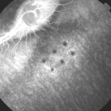
    After laser-induced choroidal neovascularization in Chinchilla rabbits, once-daily DWRX2008 drops reduced fluorescein leakage by day 14 to the same degree as a single intravitreal aflibercept injection.

    S5

    Suppression of vascular leakage in laser-induced CNV rabbits

    Chinchilla rabbit · n = 5/group · CNV & baseline day 0 → FFA day 7 → final dose & FFA day 14

    GroupCNVTreatmentRouteEyeFrequency
    G1NSaline——TID
    G2YVehicleTopicalRQD
    G3YAflibercept (Eylea®)IVT injectionRSingle
    G4YDWRX2008TopicalRQD
    Fluorescein intensity vs vehicle

    % of vehicle (G2); mean ± SD

    −28%DWRX2008 at day 14 (p<0.001); Eylea® −33%
    Fundus fluorescein angiography

    Laser spots and leakage in the choroid

    Day of CNV induction = day 0. Mean ± SD. *** p<0.001 vs G2 (vehicle). G4 = DWRX2008 high dose.

    06Safety

    Clean in the eye, proven in the body.

    GLP studies found no ocular irritation or safety findings at clinically intended and higher doses — and enavogliflozin already has a full approved-drug safety package behind it.

    DWRX2008 · topical · GLP

    • Eye irritation study

      NZW rabbits

      ✓ No findings
    • Skin irritation study

      NZW rabbits

      ✓ No findings
    • Phototoxicity

      BALB/3T3 clone A31 cells

      ✓ No findings
    • 4-week repeated topical dose toxicity, 2-week recovery, with TK

      NZW rabbits · systemic safety

      ✓ No findings

    Envlo® · oral enavogliflozin · Phase 3 ENHANCE-M

    Add-on to metformin in T2DM: no hypoglycemia

    Enavogliflozin 0.3 mg (n=101)Dapagliflozin 10 mg (n=99)

    • Patients with TEAEs
      23.8%
      22.2%
    • Patients with ADRs
      1.0%
      7.1%
    • Hypoglycemia
      0
      1.0%
    • Patients with SAEs
      1.0%
      1.0%

    Bars scaled to 25% of patients. Other ADRs with dapagliflozin: dyspepsia, pollakiuria, vulvovaginal pruritus, pruritus (1–2 patients each); cystitis in one patient per arm.

    • Complete preclinical and clinical data on Envlo® are available to expedite DWRX2008 development.

    07Where it fits

    Six ways in — alone or beside anti-VEGF.

    The last two eyedrops to try — OCS-01 and nesvategrast — both missed their endpoints. DWRX2008 — the first-in-class SGLT2 inhibitor eyedrop — is the only non-injectable candidate aimed at both the upstream (diabetic) and downstream (edema) drivers of DR/DME.

    Diabetes≥10 yrs
    NPDRnon-proliferative
    PDRproliferative
    DMEmacular edema
    Vision loss 
    1. 1

      Prevent NPDR / DME in high-risk patients

      Delay the first visible damage in long-standing diabetes.

      Diabetes → NPDR

    2. 2

      Prevent progression to PDR

      Hold patients in the non-proliferative stage, where there is generally no treatment today.

      NPDR → PDR

    3. 3

      Extend anti-VEGF dosing intervals

      Fewer injections for patients on VEGF traps.

      DME · with anti-VEGF

    4. 4

      Rescue poor anti-VEGF response

      Ischemia or occlusion, VEGF overproduction and fluctuating retinal fluid all blunt VEGF traps.

      DME · with anti-VEGF

    5. 5

      Bridge off anti-VEGF safely

      A temporary substitute that lets excessive NV inhibition or side effects unwind before re-dosing.

      DME · in place of anti-VEGF

    6. 6

      Combine in late stages

      Add a cause-level mechanism to prevent blindness.

      Late DME / PDR

    DWRX2008 compared with other treatment types
    PropertyAnti-VEGF antibodyDWRX2008 eyedropOther eyedropsSteroids
    MechanismPowerful, fast on angiogenesis; limited to downstream VEGF, weaker under ischemiaDirect anti-diabetic effect plus a second mechanism restoring endothelial function; slower on angiogenesis than VEGF trapsMostly kinase or growth-factor receptor targets, more chance of TEAEsRapid, broad anti-inflammatory; short-term use only
    RouteIntravitreal / port deliveryTrans-scleral / conjunctival (topical)Trans-corneal (topical)Intravitreal implant
    Ocular safetyRetinal detachment, IOP ↑, infection, cataractNo irritation or toxicity at intended doses (nonclinical)Off-target effects, e.g. corneal keratitisIOP ↑, cataract, hemorrhage, eye pain
    Systemic safetyGI events, hypertension, pneumonia, kidney failure, thrombosisOral form: reversible genital infection; no hypoglycemiaSimilar to anti-VEGF / TKIsImmune suppression, hyperglycemia, bone loss
    DosingEvery 1–4 monthsBID–TID dropsVariesVaries
    Cost / convenienceHigh / lowLow / highLow / highVaries
    Competitive landscape, October 2026
    ProductCompanyStatus · Oct 2026RouteMoADiabetesAngiogenesisMacular edema
    Non-invasive · eyedrop or oral
    DWRX2008Daewoong Therapeutics · Daewoong Pharmaceutical · HanAll BiopharmaPhase 1First patient in Feb 2027TopicalSGLT2 inhibitor
    First-in-class
    ●TBD●
    OCS-01 (dexamethasone)OculisPhase 3 missedDIAMOND 1 & 2 missed BCVA endpoint (May 2026); no DME filing plannedTopicalCorticosteroid——●
    Nesvategrast (OTT166)OcuTerraPhase 2 missedDR:EAM missed primary & key secondary endpoints (Mar 2024)TopicalIntegrin inhibitor——●
    APX3330Opus GeneticsPhase 3-readyFDA SPA agreed (Dec 2024); Phase 2 ZETA-1 missed primary; partner soughtOralAPE1/Ref-1 inhibitor—●TBD
    PAN-90806PanOpticaPhase 1/2Studied in wet AMD; no public update since 2020TopicalVEGFR inhibitor—●TBD
    Injected or implanted
    Vorolanib insert (Duravyu™)EyePointPhase 3COMO/CAPRI in DME fully enrolled (Jul 2026); data Q4 2027IVT insertVEGFR inhibitor—●●
    Aflibercept (Eylea®, Eylea HD®)RegeneronLaunched8 mg approved for DME & DR (Aug 2023)IVT injectionAnti-VEGF trap—●●
    Faricimab (Vabysmo®)RocheLaunchedDMEIVT injectionAnti-VEGF / Ang-2—●●
    Ranibizumab (Lucentis®)RocheLaunchedDME, DRIVT injectionAnti-VEGF mAb—●●
    Ranibizumab PDS (Susvimo®)RocheLaunchedDME (Feb 2025) and DR (May 2025) addedRefillable port implantAnti-VEGF mAb—●●
    Bevacizumab (Avastin®)RocheOff-labelNot approved for ocular useIVT injectionAnti-VEGF mAb—●●
    Dexamethasone (Ozurdex®)AbbVieLaunchedDMEIVT implantCorticosteroid——●
    Fluocinolone acetonide (Iluvien®)ANI PharmaceuticalsLaunchedDMEIVT implantCorticosteroid——●

    Competitive landscape updated October 2026 from company announcements and trade press (Oculis, OcuTerra, Opus Genetics, EyePoint, Regeneron, Roche). Diabetes / angiogenesis / macular edema columns show which disease drivers each mechanism targets.

    08Clinical plan

    Into the clinic: first patient in February 2027.

    Phase 1 starts with first patient in planned for February 2027. With full systemic data from the approved oral drug behind it, DWRX2008 is set up to move quickly into a Phase 2 efficacy study in DME patients.

    Phase 1 · safety, tolerability, PK

    Design
    Parallel, randomized, double-blind, placebo-controlled; single and multiple ascending application
    Subjects
    60 healthy Korean and Caucasian volunteers · 6 SAD and 3 MAD cohorts
    Assess
    Local safety (irritation, redness), ophthalmic exams, PK, urinary glucose excretion, insulin, HbA1c, fasting glucose
    Timing
    First patient in planned for February 2027

    Concentrations tested

    0.5%
    1%
    2%
    3%

    2% BID–TID is the intended clinical dose.

    Path forward

    1. Done

      Nonclinical package

      In vitro pharmacology, ocular PK and QWBA, two efficacy models, GLP irritation, phototoxicity and 4-week repeated-dose toxicity.

    2. Feb 2027

      Phase 1 first patient in

      SAD/MAD design in healthy volunteers.

    3. Next

      Long-term repeated-dose toxicity (GLP)

      With existing systemic data, enables a direct move to Phase 2.

    4. Next

      US FDA pre-IND → Phase 2 IND

      Efficacy study in DME patients; cGMP manufacture of Phase 2 supplies.

    5. Partnering

      Milestone-based license-out or co-development

      Partnering alongside the three-company development team.

    09Co-development

    Three companies, one program — proven in US ophthalmology.

    DWRX2008 is co-developed by Daewoong Therapeutics, Daewoong Pharmaceutical and HanAll Biopharma. Both partners have already run US Phase 3 trials of an eyedrop in dry eye disease.

    Formulation & delivery

    Daewoong Therapeutics

    • Originator of DWRX2008 and its nano-micelle eyedrop formulation
    • Drug delivery R&D specialist; holds the ophthalmic-use rights
    • Built the nonclinical package: pharmacology, ocular PK, efficacy, GLP safety

    API & global development

    Daewoong Pharmaceutical

    • Developer of enavogliflozin, approved in Korea as Envlo® — a full preclinical and clinical package behind DWRX2008
    • Global pharmaceutical company with regulatory and commercial reach
    • Co-ran the US Phase 3 VELOS program of tanfanercept eyedrops in dry eye disease with HanAll

    US Phase 3 · dry eye

    Ophthalmic clinical development

    HanAll Biopharma

    • Developer of tanfanercept, a TNF-α inhibitor eyedrop for dry eye disease
    • Ran the US Phase 3 VELOS program (VELOS-3, VELOS-4) with Daewoong Pharmaceutical
    • Hands-on experience with US eyedrop trial design, sites and endpoints

    US Phase 3 · dry eye

    2 of 3

    partners have already taken an eyedrop into US Phase 3 — validated ophthalmology partners from day one

    Eyedrop

    Same dosage form, same US trial know-how: ocular tolerability, ophthalmic exams, retina and cornea sites

    API → eye

    An approved SGLT2 API, a delivery platform and an ophthalmic clinical team in one program

    Bring a drop to the back of the eye.

    DWRX2008 is open to licensing and joint development. Talk to us about the full data package.

    Partner on DWRX2008

    DWRX2008 is an investigational product and is not approved for any indication. Nonclinical results do not predict clinical outcomes. Charts are redrawn from Daewoong Therapeutics study reports; values read from source figures are approximate. Lucentis®, Eylea®, Avastin®, Vabysmo®, Susvimo®, Ozurdex® and Iluvien® are trademarks of their respective owners. Envlo® is a trademark of Daewoong Pharmaceutical. Tanfanercept is an investigational product of HanAll Biopharma.